Explainable identification of lung abnormalities in chest X-ray and CT imaging
While the use of machine learning/AI for medical image interpretation has seen tremendous growth in the research domain over several years, clinical translation of developed solutions remains limited. This may at least in part be attributed to the simplification of predictive tasks, when developing ML/AI methods. For example, detection and identification of lung abnormalities in chest imaging are often posed as binary or multi-class classification problems wherein, a few types of abnormalities (e.g. pneumonia, nodules, pleural effusion, etc.) are selected as the targets of interest for the predictive task. This ignores the true underlying challenge in the predictive task in that abnormalities often co-occur, especially in populations at high risk of respiratory diseases. In doing so, developed solutions have limited clinical relevance and applicability.
To address this limitation, researchers at the University of Leeds, led by Rachael Harkness (PhD student, School of Computing) and Nishant Ravikumar (Lecturer, School of Computing), developed a ‘multi-label’ classification approach to tackle the challenging and clinically relevant problem of predicting the presence of multiple co-occurring lung abnormalities in chest X-ray images (presented at the SPIE Medical Imaging 2023 conference). The developed method is based on disentangled representation learning using Dirichlet Variational Autoencoders, which results in an inherently explainable predictive model. Specifically, the learned sparse, disentangled latent space is highly correlated with the target abnormalities observed in the CheXpert dataset, allowing for ‘latent traversals’ which enable localisation of the abnormalities as a by-product of the multi-label classification task (see Figure 1). The developed approach is currently being expanded to the problem of unsupervised anomaly detection and identification of co-morbidities alongside lung abnormalities, using chest CT imaging.
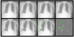
Figure 1:Automatic identification of lung opacity using latent traversals
A recently funded project by Leeds Hospital Charity will enable the deployment of the developed approach to analyse GP referred chest X-ray images from 70,000 patients, collected at Leeds Teaching Hospitals, to identify patients at risk of lung cancer within a two-year period. The overarching goal of this project is to demonstrate the clinical applicability of the developed approach for chest X-ray interpretation and to assess its performance compared to radiologist reporting.