Meta-analysis of the normal diffusion tensor imaging values of the peripheral nerves in the upper limb

Peripheral neuropathy affects 1 in 10 adults over the age of 40 years. Given the absence of a reliable diagnostic test for peripheral neuropathy, there has been a surge of research into diffusion tensor imaging (DTI) because it characterises nerve microstructure and provides reproducible proxy measures of myelination, axon diameter, fibre density and organisation.
Before researchers and clinicians can reliably use diffusion tensor imaging to assess the ‘health’ of the major nerves of the upper limb, we must understand the “normal” range of values and how they vary with experimental conditions. We searched PubMed, Embase, medRxiv and bioRxiv for studies which reported the findings of DTI of the upper limb in healthy adults.
Four review authors independently triple extracted data. Using the meta suite of Stata 17, we estimated the normal fractional anisotropy (FA) and diffusivity (mean, MD; radial, RD; axial AD) values of the median, radial and ulnar nerve in the arm, elbow and forearm. Using meta-regression, we explored how DTI metrics varied with age and experimental conditions.
We included 20 studies reporting data from 391 limbs, belonging to 346 adults (189 males and 154 females, ~ 1.2 M:1F) of mean age 34 years (median 31, range 20–80).
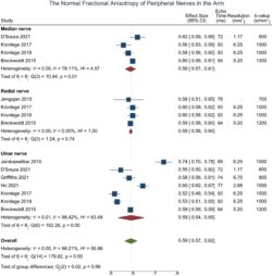
In the arm, there was no difference in the FA (pooled mean 0.59 mm2/s [95% CI 0.57, 0.62]; I2 98%) or MD (pooled mean 1.13 × 10⁻³ mm2/s [95% CI 1.08, 1.18]; I2 99%) of the median, radial and ulnar nerves.

Figure 1 - The normal fractional anisotropy of peripheral nerves in the arm
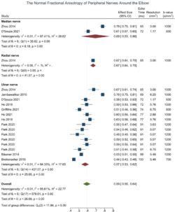
Around the elbow, the ulnar nerve had a 12% lower FA than the median and radial nerves (95% CI − 0.25, 0.00) and significantly higher MD, RD and AD.

Figure 2 - The normal fractional anisotropy of peripheral nerves in the elbow
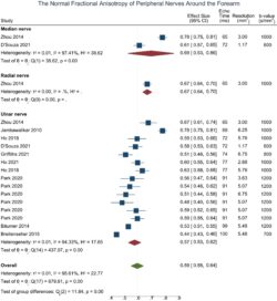
In the forearm, the FA (pooled mean 0.55 [95% CI 0.59, 0.64]; I2 96%) and MD (pooled mean 1.03 × 10⁻³ mm2/s [95% CI 0.94, 1.12]; I2 99%) of the three nerves were similar.

Figure 3 - The normal fractional anisotropy of peripheral nerves in the forearm
Multivariable meta regression showed that the b-value, TE, TR, spatial resolution and age of the subject were clinically important moderators of DTI parameters in peripheral nerves. We show that subject age, as well as the b-value, TE, TR and spatial resolution are important moderators of DTI metrics from healthy nerves in the adult upper limb. The normal ranges shown here may inform future clinical and research studies.
